
Daily Podcast full article
Medical AI Reads Faces and Scans for Hidden Health Signals
Two new medical AI reports point in the same direction: routine images may contain more clinical information than doctors currently extract. One model estimates biological age and cancer survival risk from a single face photograph, while another, Percival, searches CT scans for disease signals beyond the reason the scan was ordered. The promise is earlier, cheaper, more remote assessment; the catch is that both systems still need rigorous validation, privacy controls and proof that their signals improve care rather than amplify bias.
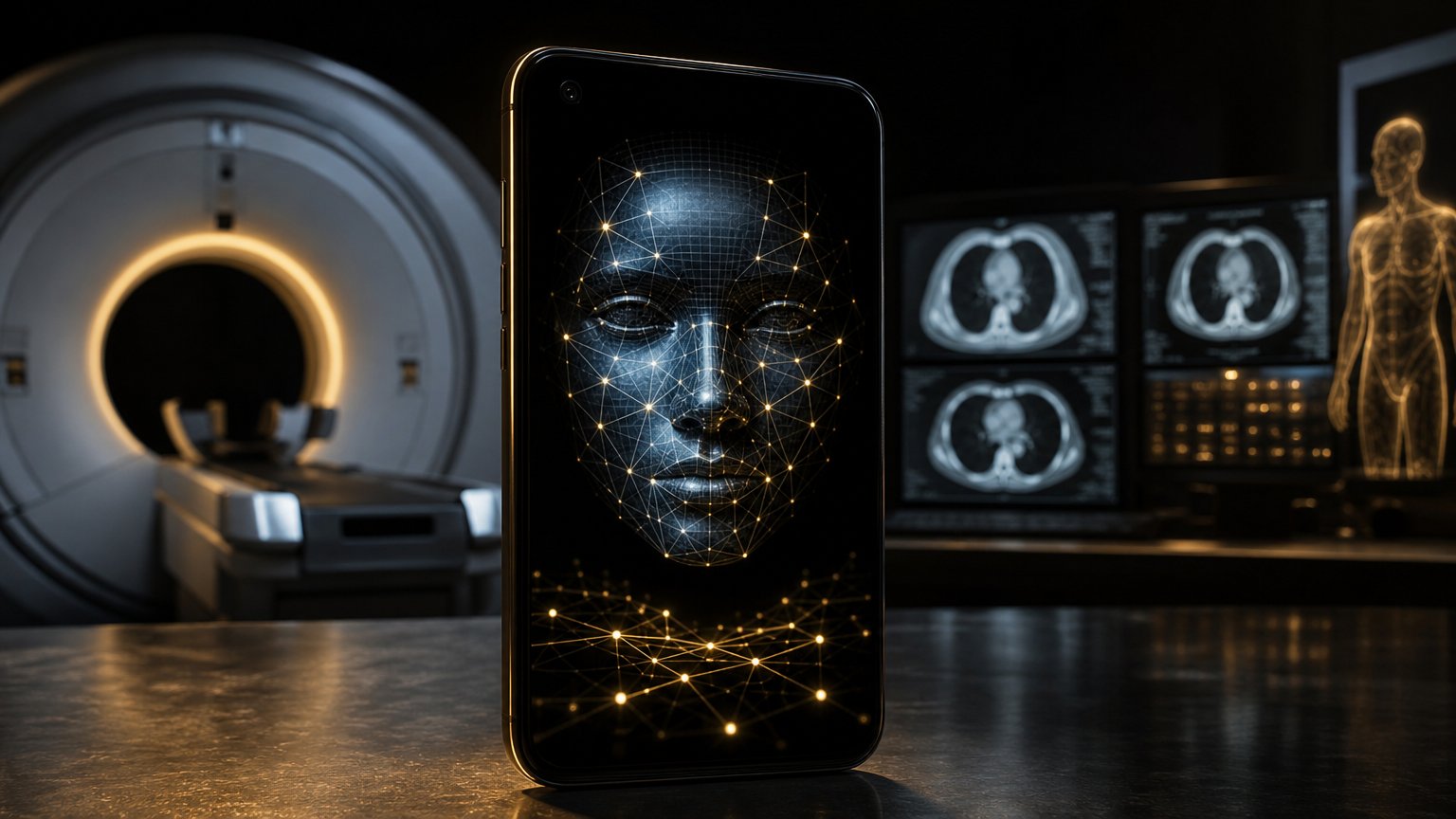
A selfie, a scan and the same clinical question
The working headline is the story: medical AI is beginning to read faces and scans for health signals that were not the original target of the image. In one line of work, Mass General Brigham researchers built FAHR-Face, a foundation model trained on more than 40 million face images and adapted into tools that estimate biological age and predict survival outcomes in patients with cancer . In the other, Penn Medicine’s Percival model analyzes CT scans to identify disease patterns and future health risks beyond the clinical question that prompted the scan .
The two systems are not the same technology and should not be treated as equal clinical products. FAHR-Face starts from ordinary facial photographs; Percival starts from three-dimensional medical imaging. But they share a larger thesis: a medical image may carry weak, distributed signals about frailty, disease burden or future risk that are visible to a large model even when they are not part of routine interpretation.
That thesis is attractive because it does not require a new biopsy, a new scanner or a rare specialist at every site. A face photograph can be captured almost anywhere, and a CT scan already exists in many hospital records. The risk is equally clear: if an AI turns a face or incidental scan pattern into a clinical score, the health system must know whether it is reading biology, socioeconomic history, camera quality, race, age, treatment access, or some mixture of all of them.
What the face model claims to do
FAHR-Face was developed as a large facial health recognition model and then used to create two related tools: FAHR-FaceAge and FAHR-FaceSurvival . FAHR-FaceAge estimates biological age from a face photograph, while FAHR-FaceSurvival predicts survival outcomes in cancer patients directly from a facial image rather than using age estimation as an intermediate step .
The Guardian reported that the system was custom-built by Mass General Brigham experts to estimate biological age and directly predict survival outcomes for cancer patients, with details published in The Lancet Digital Health . The Straits Times reported that the model used more than 40 million face images and more than 30,000 cancer patient photographs, and that its age estimates were off by about five years . Those figures matter because they frame the tool as a population-scale risk model, not a magic diagnostic camera.
The most clinically interesting idea is that appearance might quantify something doctors already notice informally. Oncologists and surgeons routinely judge whether a patient appears robust enough for chemotherapy, radiotherapy, surgery or a combined regimen. A model that turns visual frailty or biological age into a consistent score could, in theory, support decisions when lab values, genomic tests or specialist evaluation are incomplete.
But “support” is the operative word. Reports on the study emphasize that the technology is not ready to replace physician judgment and still needs testing across hospitals and patient groups . That caveat is not a footnote; it is the central condition for usefulness. If the score performs well only in a narrow set of patients or camera conditions, it could create false reassurance for some groups and false alarm for others.
Why bias and privacy are not side issues
Face-based medicine sits directly on top of one of AI’s most sensitive data categories: biometrics. A facial photograph can identify a person, reveal age, sex presentation and ancestry cues, and encode social determinants of health. If a model predicts survival partly because facial features correlate with unequal access to nutrition, dental care, housing, sun exposure, stress or prior treatment, then the model may be detecting real risk while also reproducing the inequities that produced it.
That does not make the research useless. It makes validation harder. A clinically responsible face model would need performance reporting by race, age, sex, cancer type, disease stage, imaging conditions and geography. It would also need consent rules that are more explicit than ordinary clinical photography, because a photo taken for identity verification or radiotherapy setup could become a prognostic input.
There is also a communication problem. A patient should not be told that an AI “read their face” and predicted survival without a careful explanation of uncertainty. A risk score is not a life expectancy. It is a probability signal derived from past data. Its proper use would be to add one calibrated piece of evidence to a clinical conversation, not to determine treatment eligibility on its own.
Percival and the hidden information inside CT scans
The CT story is less emotionally charged than facial analysis, but it may be just as important operationally. Penn Medicine’s Percival model was trained on roughly 400,000 CT scans from about 50,000 patients, using data connected to the Penn Medicine BioBank, which includes information from more than 350,000 people collected across the University of Pennsylvania Health System over two decades .
According to Newswise’s account of the Penn release, Percival can uncover nearly hidden disease patterns and potential future health risks that may otherwise go unnoticed by human readers, and it can run in the background rather than being prompted to find only one condition . Medical Xpress reported that the model found traces of chronic conditions including atrial fibrillation and chronic kidney disease, and that it flagged pancreatic cancer, cirrhosis and bacterial pneumonia more reliably than TotalSegmentator in the reported comparison .
The concept is often called opportunistic screening: using an image acquired for one reason to find additional clinically relevant information. A chest or abdominal CT ordered for pneumonia, trauma or pain may contain information about vascular calcification, organ size, body composition, kidney disease, liver disease or early malignancy. A model that can systematically extract such signals could make existing scans more valuable without exposing patients to more radiation.
That value proposition is especially relevant to cost. CT scans are expensive, and repeat imaging adds radiation exposure, scheduling friction and follow-up burden. If an AI can extract more actionable information from a scan that has already been performed, hospitals could in principle detect risk earlier without ordering a new study.
The deployment bar should be high
Percival is still being tested preclinically, and the Penn team is looking toward further testing before regular clinical use . News-Medical likewise described the model as a new AI system for detecting hidden diseases in routine CT scans, but the current reports should be read as research progress, not a cleared clinical service .
The missing pieces are familiar but decisive. Clinicians need sensitivity, specificity, positive predictive value, calibration, external validation and workflow studies. They also need to know how many false positives the model creates. Opportunistic screening can save lives when it finds real disease early, but it can also trigger anxiety, extra imaging, biopsies and downstream procedures for findings that would never have harmed the patient.
That is why “the model saw something” is not enough. A hospital needs to know whether acting on that something improves outcomes. Does Percival find pancreatic cancer early enough to change survival? Does it identify chronic kidney disease before standard labs would? Does it help cardiologists prevent complications, or merely label patients who were already visibly ill in the electronic health record?
The bigger pattern: ambient prognosis
Taken together, FAHR-Face and Percival suggest a future of ambient prognosis: health systems using images already collected in care to infer risk in the background. The selfie or clinic photo becomes a rough proxy for biological age; the CT becomes a broad health map rather than a single-purpose exam.
That future could help rural hospitals, under-resourced clinics and remote oncology programs if it is deployed carefully. A low-cost facial risk signal might help prioritize specialist review. A CT foundation model might alert physicians to disease that was outside the original radiology request. Both could make medical assessment more continuous and personalized.
But the same future could also normalize surveillance, opaque triage and automated pessimism. The line between a useful early warning and a biased gatekeeping score will depend on evidence, governance and patient consent. The technology is moving quickly; the clinical proof must move just as carefully.
For now, the story is not that AI can diagnose everything from a selfie or a scan. The story is narrower and more interesting: researchers are finding that ordinary medical images may carry measurable signals about age, disease burden and survival. If those signals hold up across diverse patients and real-world workflows, medicine may get a new class of non-invasive prognostic tools. If they do not, the field will have learned again that seeing a pattern is easier than proving it should guide care.
Sources from the last 72 hours
- [1]AI Facial Scans Help Doctors Assess Age and Survival RiskOct 20, 2026, 12:45 AM
- [2]New AI tool can predict cancer survival by a single patient selfie, study suggestsOct 20, 2026, 12:30 AM
- [3]AI tool predicts cancer survival chances with single face photo, study findsOct 20, 2026, 1:50 AM
- [4]AI Model Can Uncover Unseen Health Issues in CT ScansOct 20, 2026, 12:25 AM
- [5]Percival AI can uncover unseen health issues in CT scansOct 19, 2026, 2:00 AM
- [6]New AI model detects hidden diseases in routine CT scansOct 20, 2026, 2:00 AM
AI-generated article based on recent web research, then preserved as a dated editorial snapshot.
Comments
Be the first to comment.