
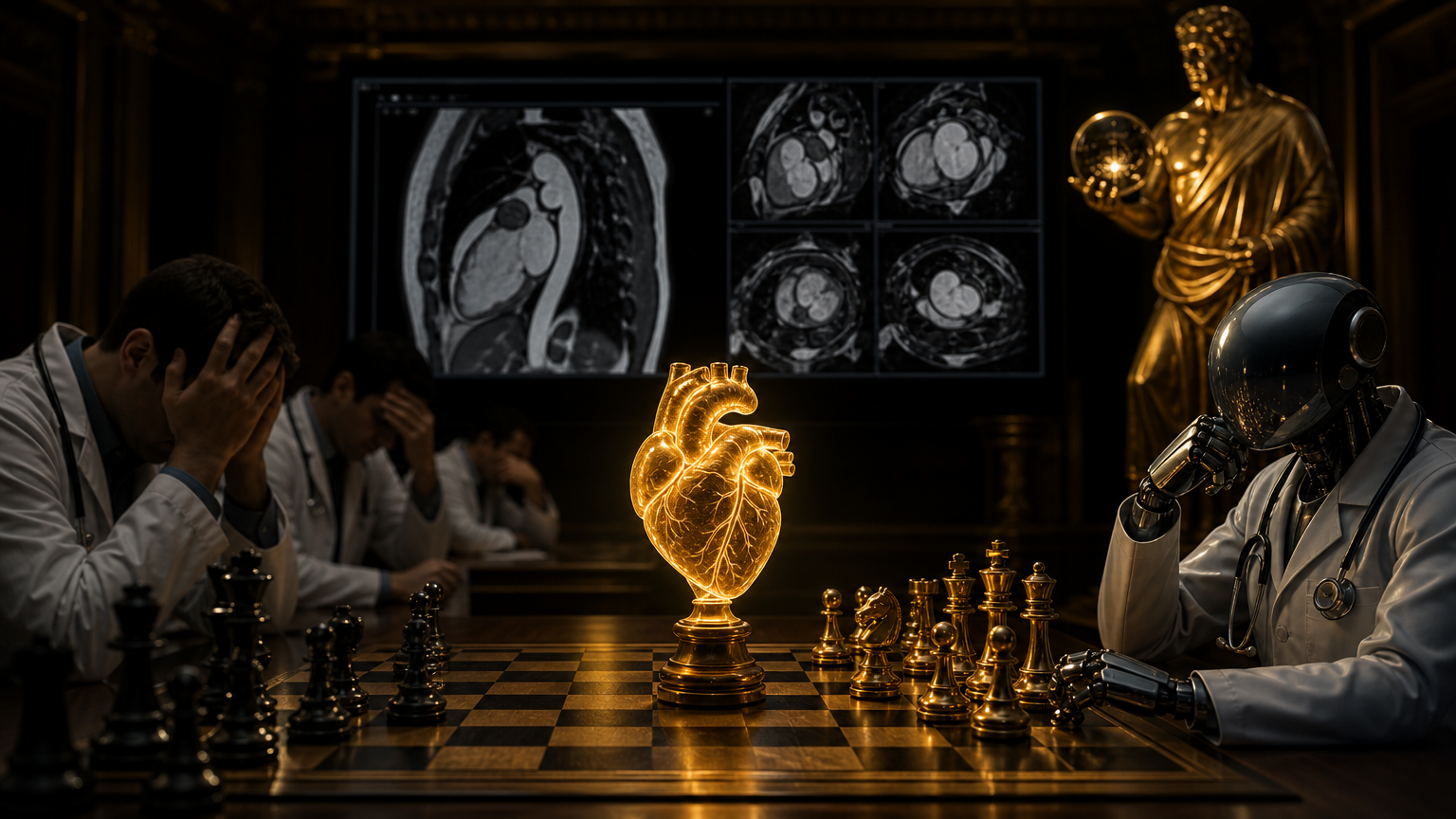
The shift: from drawing borders to predicting survival
The headline in this study is not simply that an AI system can outline the left ventricle on a cardiac MRI. Many imaging algorithms already perform segmentation, measurement, or detection tasks. The more important shift is that the resulting measurement was tested against a clinical outcome that hospitals and patients understand immediately: survival.
Researchers led by City St George’s, University of London, with University College London collaborators, reported that AI measurements of heart function from MRI scans were more strongly linked to how long patients lived after their scan than equivalent measurements made by clinicians . The work, published in The Lancet Digital Health, focused on ejection fraction, the proportion of blood the left ventricle pumps out with each beat and one of cardiology’s most consequential measurements .
Ejection fraction is not an obscure research variable. It is used to guide treatment decisions for heart disease, including specialist heart failure therapies, implanted heart devices, procedures to improve blood flow, surgical risk assessment, and monitoring of cardiac function during chemotherapy . That is why even a modest improvement in how the measurement relates to outcomes matters. In this setting, the AI was not being judged by whether it matched a human label; it was judged by whether its measurement better tracked mortality.
What the researchers studied
The study analyzed cardiac MRI scans from 27,382 patients undergoing investigation for known or suspected heart conditions in the United Kingdom and the United States . Patients were followed for an average of more than five years, during which 3,458 deaths occurred . Truvace’s summary of the study describes the design as a retrospective, multicenter prognostic study and notes that the cohort included 26,106 patients from Barts Heart Centre and 1,276 from the University of Pittsburgh Medical Centre .
The comparison was direct: ejection fraction generated by a deep-learning AI segmentation tool versus ejection fraction produced by experienced clinicians assessing the same cardiac MRI scans . Both were then tested for association with all-cause mortality . This distinction is important. The AI was not asked to forecast death from a black box of every available patient variable; it was asked to measure heart-pumping function from MRI in a way that better stratified risk.
According to the institutional release, AI-derived ejection fraction was slightly but significantly better at identifying patients at increased risk of death than clinician-derived ejection fraction, and the finding remained significant after accounting for age and sex . Truvace’s review reports that AI ejection fraction had better model fit and marginally better discrimination than clinician ejection fraction, including an unadjusted C-index of 0.669 versus 0.637 and an age-and-sex-adjusted comparison of 0.781 versus 0.777 .
Those numbers should be read carefully. This is not a giant performance leap. It is a small statistical advantage in a large dataset. But in cardiology, a small gain attached to a routine gatekeeper measurement can still affect many decisions when applied across tens of thousands of scans.
Speed is the operational story
The survival signal is the scientific story. The workflow story is speed. The researchers reported that the AI tool analyzed scans in roughly 2–3 seconds per patient, compared with earlier work from the team showing that expert clinician analysis typically takes about 13 minutes . News-Medical’s report repeats the same comparison and frames the tool as a possible first-line approach to measuring heart function .
That speed difference matters because cardiac MRI is valuable but resource-intensive. A hospital does not need AI only because it is curious about automation; it needs AI if automation can remove bottlenecks without weakening quality. In this case, the proposed role is not “AI signs the report and sends the patient home.” It is closer to “AI performs the first measurement quickly, flags the quantified result, and leaves clinicians to verify, contextualize, and decide.”
The study authors and university reports repeatedly emphasize clinician oversight . That caveat is not decorative. Cardiac imaging measurements are used in high-stakes pathways: defibrillator consideration, heart failure treatment escalation, revascularization decisions, perioperative risk discussions, and chemotherapy surveillance. A measurement can be numerically precise and still clinically misleading if the scan quality is poor, the anatomy is unusual, or the broader patient context points elsewhere.
The error profile still needs governance
The release states that AI errors were uncommon, with an error rate of around 1% at the patient level affecting just 0.08% of images . That sounds reassuring, and in operational terms it is. But a 1% patient-level error rate in a large imaging service is not zero. The larger the deployment, the more rare errors become routine events.
This is why the most plausible adoption pathway is supervised automation. The AI can measure every scan quickly and consistently, while experts review cases where the output looks implausible, where the image quality is borderline, or where the result will trigger major treatment decisions. The clinical question is not whether AI makes any mistakes. It is whether its errors are detectable, whether its benefits outweigh those errors, and whether accountability is clear when the measurement enters the medical record.
The authors’ own interpretation is measured rather than triumphalist. Truvace summarizes the study as supporting potential utility for first-line ejection-fraction measurement with clinician oversight, while noting that prospective studies are still needed to confirm practical benefit . That is the correct posture. Retrospective multicenter evidence can show that an AI-derived measurement correlates better with survival than a clinician-derived measurement. It cannot, by itself, prove that deploying the tool tomorrow will improve patient outcomes.
Why survival prediction changes the AI debate
Medical AI often begins with label replication: Can the system contour the chamber? Can it detect a lesion? Can it classify a scan like a specialist? Those are necessary steps, but they can leave hospitals with an uncomfortable question: even if the AI matches the label, does that make patients better off?
This study pushes the benchmark toward outcomes. If an AI measurement predicts mortality more consistently than the conventional human-derived value, hospitals can ask more concrete questions. Does the tool move high-risk patients into earlier follow-up? Does it reduce waiting time for reports? Does it improve consistency across sites and readers? Does it help identify borderline patients who might otherwise be under-monitored?
The research team is already pointing beyond MRI-only measurement. The City St George’s and UCL releases say the team plans to combine AI measurements from heart scans with other tests, particularly electrocardiograms, to identify heart muscle problems earlier and allow treatment before lasting damage occurs . That next step matters because single-measurement AI may become most powerful when integrated into a longitudinal risk picture: imaging, ECGs, laboratory values, symptoms, medications, and outcomes.
What hospitals should watch next
For hospital leaders, the study creates a useful but demanding checklist. First, external validation must confirm performance beyond the study settings and scanner mixes. Second, workflow integration must be tested in real reporting environments, not only in retrospective archives. Third, clinicians need clear thresholds for when to accept, review, or override the AI output. Fourth, governance teams need audit trails: which version of the model measured the scan, whether a human reviewed it, and how discrepancies were resolved.
The study also gives buyers a better way to judge cardiac imaging AI. Vendors often promise faster reads, reduced workload, or consistency. Survival-linked performance is a harder and more meaningful benchmark. If a system’s measurement better predicts mortality, it moves from convenience technology toward risk-stratification infrastructure.
The right takeaway is neither “AI beats doctors” nor “AI is just a faster ruler.” The better reading is that, in this large cardiac MRI cohort, an AI-derived ejection fraction carried slightly more prognostic information than the clinician-derived version and did so far faster . That is a real advance, but it is also a supervised one. The tricorder may be submitting peer-reviewed benchmarks now, but cardiology still has to decide how those benchmarks become safer care.
Comments
Be the first to comment.